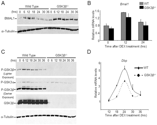
Figure 4. Circadian defects in GSK3β−/− MEFs.
WT and GSK3β−/− MEFs were synchronized by 2 hour treatment with 100nM dexamethasone (DEX). (A) Total lysates were prepared at indicated times post synchronization and resolved by SDS-PAGE followed by Western analysis. BMAL1 and α-tubulin levels were detected by specific antibodies. (B) RNA was prepared at indicated times, reverse transcribed, and real-time PCR was performed using primers for Bmal1 and 18S rRNA. Data is represented as relative levels of Bmal1 normalized to 18S rRNA. (C) Same as in (A), except phospho-GSK3α/β and total GSK3β levels were detected by specific antibodies. (D) RNA was prepared at indicated times, reverse transcribed, and real-time PCR was performed using primers for Dbp and 18S rRNA. Data is represented as relative levels of Dbp normalized to 18S rRNA.

