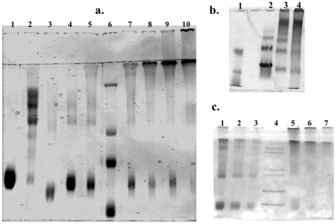
Figure 1. Analysis of bovine serum albumin (BSA) samples by polyacrylamide gel electrophoresis (PAGE).
Gels are 7.5% polyacrylamide (PA) with stacking gel of 2% PA and stained with Coomassie Blue. (a) SDS-PAGE: Samples were incubated at 95° C in denaturing sample buffer (containing β-mercaptoethanol and SDS), and each lane was loaded with 10 mg of protein and run with SDS in the buffer. Lane 1: BSA monomer; lane 2: BSA dimer; lane 3: methylated BSA (Sigma Chemicals); lanes 4, 5, 7, 8, 9, 10 contain 10% BSA monomers crosslinked with increasing [GA]: 0.1%, 0.2%, 0.3%, 0.4%, 0.5%, 0.6% GA respectively; lane 6: molecular weight marker (229,126,80,48 kDa). (b) Non-denaturing PAGE. Lane 1: molecular weight marker (85, 50, 35 kDa); lane 2,3,4 are fraction V BSA with 0.0%, 0.4%, and 0.5% GA respectively. (c) Non-denaturing PAGE: Lanes 1–3: methylated BSA (Sigma Chemicals) with decreasing amounts (5, 2.5 and 1.25 ug) of loaded protein; lane 4: molecular weight marker (200, 116, 97, 66, 55 kDa); lanes 5–7: 10% fraction V BSA reacted with 0.4% GA in decreasing amounts (5, 2.5, and 1.25 ug) of protein loaded. See text for details.

