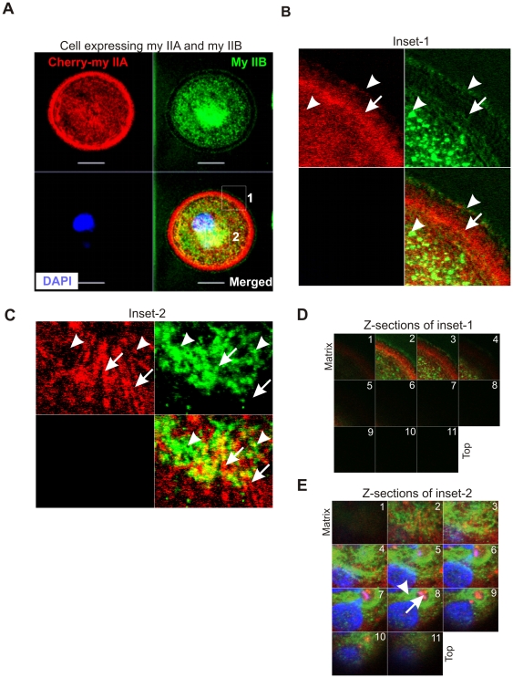
Figure 3. Distinct localizations of myosin IIA and IIB in spreading MDA-MB 231 breast cancer cells.
A) Spreading cell transiently expressing cherry-myosin IIA was fixed and stained with myosin IIB antibody. A series of Z-sections were collected using confocal microscope. Inset-1 and inset-2 were drawn to study colocalizations of myosin IIA and IIB at the spreading margins and in the cytoplasm, respectively. B) and C) Blowup of a z-sections collected near the matrix. Arrow indicates cherry-myosin IIA and arrow-head shows endogenous myosin IIB. D) and E) Myosin IIA and IIB display distinct localization in the spreading margins and cytoplasm. A series of z-sections were collected using confocal microscope.

