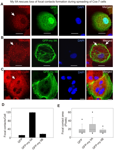
Figure 7. Transient expression of GFP-myosin IIA but not IIB rescues loss of focal contacts formation in spreading COS-7 cells.
Cells were fixed after 60–70 min of spreading and stained with vinculin antibodies and DAPI. A) Loss of focal contacts in spreading COS-7 cells transiently expressing GFP. Arrow indicates loss of focal contact formation in the spreading cell. B) Transient expression of GFP-myosin IIA rescues formation of focal contacts. Arrow indicates focal contact formation. C) Transient expression of GFP-myosin IIB does not rescue focal contacts formation in the spreading cells. Arrow indicates loss of focal contact formation. D) Graphical representation of number of punctate structures (focal contacts) per cell. Confocal z-sections were exported into TIF images and then processed to quantify number and area of punctate structures using ImageJ program (NIH). Numbers of images processed to quantify number and area of punctate structures were in between 12–15. E) Box-and-whisker plot showing the average area of focal contact. We have observed a wide range of areas of punctate structures in pixels and therefore showed by Box-and-whisker plot.

