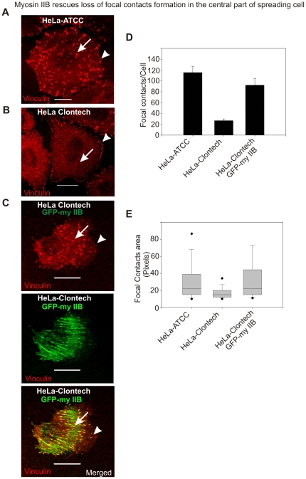
Figure 8. Myosin IIB is required for focal contacts formation in the central part of the spreading cells.
A) Spreading HeLa-ATCC cell displaying focal contacts formation through out the membrane. Arrow and arrow-head indicate focal contacts formation in the central and cell margins of the spreading cell, respectively. B) Spreading HeLa-Clontech cells show focal contacts formation exclusively in the peripheral region. Arrow indicates loss of focal contacts formation in the central region and arrow-head shows the formation of focal contacts in the spreading margins. C) Transient expression of GFP-myosin IIB in HeLa-Clontech cells rescues the formation of focal contacts in the central part of spreading cell. The scale bars represent 20 µm. D) Graphical representation of focal contacts in spreading HeLa cells. E) Box-and-whisker plot showing the average area of focal contact. Numbers of images processed to quantify number and area of punctate structures were in between 12–16.

