
Fig 5.
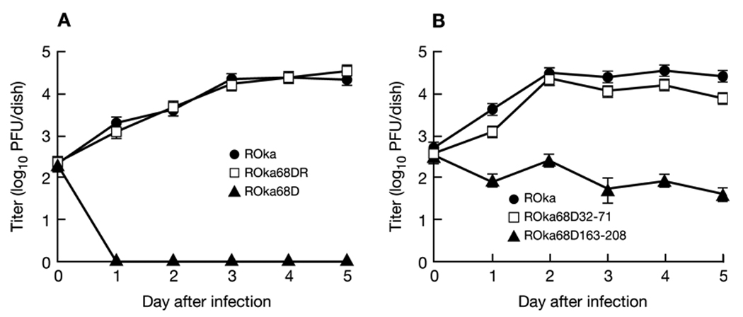
Growth of VZV gE deletion mutants. Melanoma cells were infected with VZV ROka, ROka68DR, or ROka68D (A), or ROka, ROka68D32-71, or ROkaD163-208 (B), and the titer of virus in cells was assayed on days 0 to 5 after infection. Each value represents the average number of plaques in two wells and error bars indicate standard errors. The experiment was performed twice and a representative result is shown.