
Fig. 9.
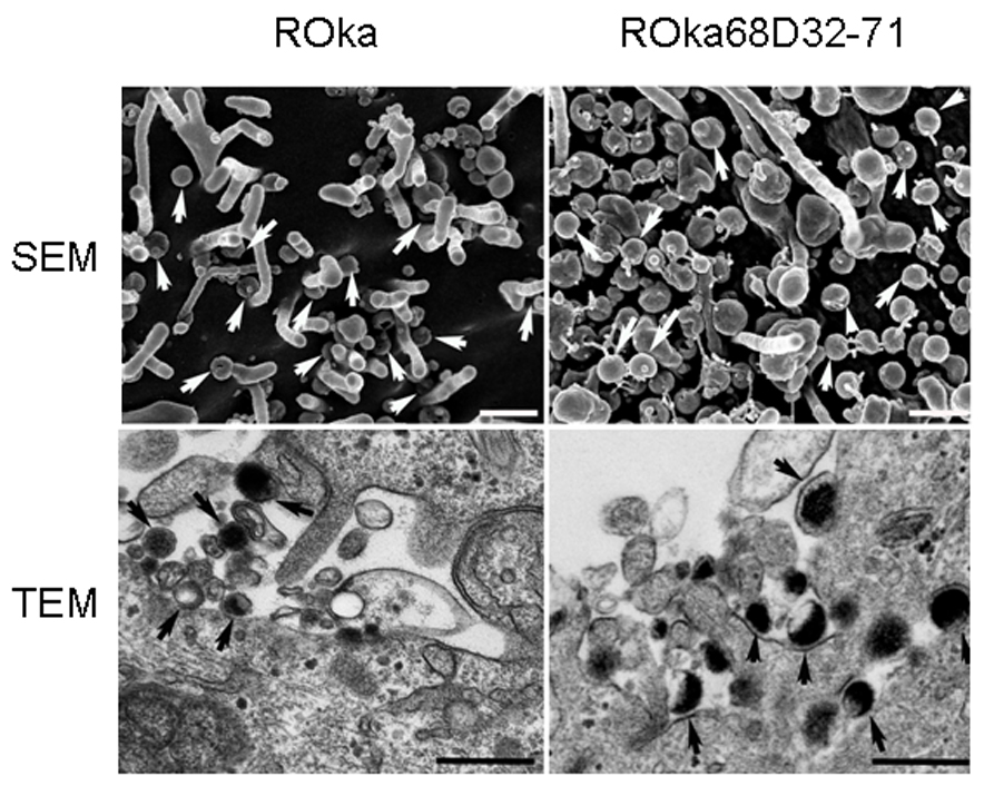
VZV deleted for the IDE binding domain of gE traffics to the cell surface. Melanoma cells were infected with VZV ROka or ROka68D32-71, cells were fixed as described in the Methods, and scanning electron microscopy (SEM) or transmission electron microscopy (TEM) was performed. Arrows indicate virions and bars are 400 nm.