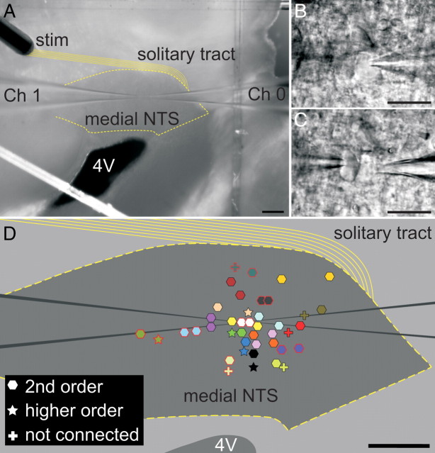
Figure 1.
The distribution of neurons recorded as pairs within the medial NTS in a horizontal slice of the caudal brainstem. A, For experiments, the concentric bipolar electrode (stim) was placed over rostral portions of the solitary tract (solid yellow lines). Two pipettes (Ch 0 and Ch 1) were independently advanced to record from cell bodies. B, C, Higher-magnification photographs of the pair of neurons in A using DIC optics and infrared imaging; we designated neurons recorded from Ch 0 as neuron “a” (B) and those recorded via Ch 1 as neuron “b” (C). D, On the basis of synaptic responses, neurons were classified as second order (hexagon), higher order (star), or not connected (cross). Both members of each NTS neuron pair are coded with like colors. The locations of the cell bodies for 21 pairs within the medial NTS are represented. Note that the locations of three pairs (3 second order, 2 higher order, and 1 not connected) are not plotted. Scale bars: A, D, 200 μm; B, C, 20 μm.

