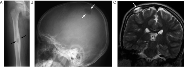
Figure 1.
An 8-year-old girl with Langerhan's cell histiocytosis (LCH) presented with right thigh pain. (A) This anterior-posterior (AP) radiograph shows a lytic lesion surrounded by cortical thickening (arrows) in the diaphysis of the right femur. The femur is a common site of involvement in LCH. (B) The patient also had a skull lesion as shown in this lateral skull x-ray (arrows). Note the beveled edges of this lesion, a feature typical of LCH. (C) This coronal T2-weighted MRI shows the soft tissue component (arrows) of the skull lesion. The skull is the most common site of bone disease in LCH and patients often present with a palpable scalp mass.

