Figure 3.
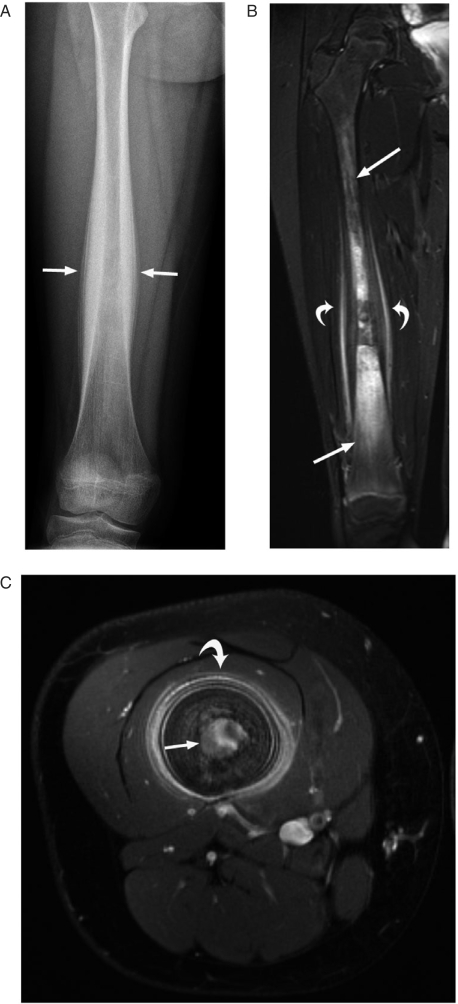
A 10-year-old girl with a 1-year history of right leg pain due to Ewing sarcoma. (A) This AP radiograph shows thick lamellated, periosteal reaction in the mid-shaft of the right femur (arrows). (B) This short tau inversion recovery (STIR) coronal MR image shows the poorly defined tumor infiltrating the marrow space (straight arrows) and thick periosteal reaction surrounded by soft tissue edema (curved arrows). (C) This axial contrast-enhanced MR image shows the enhancing marrow tumor (straight arrow) and thick, lamellated, periosteal reaction (curved arrow). The mid-shaft location and lamellated periosteal reaction are features typical of Ewing sarcoma.

