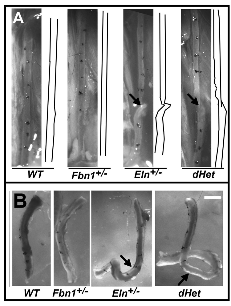
Figure 1. Morphology of ascending aortae and left common carotid arteries.
Panel A: Representative left common carotid artery of WT, Fbn1+/−, Eln+/−, and dHet mice with the tracing of vessels shown on the right side of each image. Arrow points to a vessel twist. Panel B: Representative carotid arteries of the indicated genotypes upon excision with the arrows highlighting arterial tortuosity. Scale bar = 1 mm.

