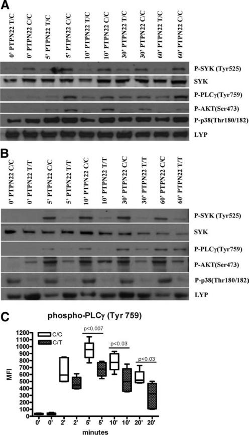
FIGURE 3.
B cells from subjects harboring the 1858 T variant display BCR-mediated signaling defects. B cells were isolated and then activated with anti-IgM for the indicated time in minutes (′), followed by cell lysis. Western blotting was performed with the following Abs: phospho (P)-Akt-Ser473, phospho-PLCγ2-Tyr759, Syk, phospho-Syk-Tyr525/526, phospho-p38 MAPK-Thr180/Tyr182, and Lyp. A, Two control subjects were compared, one homozygous for 1858C/C and the other heterozygous for 1858C/T. B, Two control subjects were compared, one homozygous for 1858C/C and the other homozygous for the1858T/T variant. Blots are representative of three independent experiments. C, B cells were stimulated with soluble anti-IgM plus IgG F(ab′)2 for the indicated times. Total phospho-PLCγ2 (Tyr759) was assessed by intracellular flow cytometry in the CD27+ memory population of 1858 C/C and 1858 C/T subjects (n = 6). Mean fluorescence intensity (MFI) values for phospho-PLCγ2 are shown.

