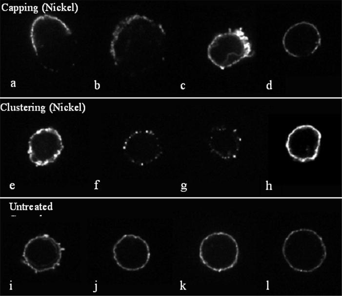
Figure 8.
Metal induced effects on T-cell receptor clustering of lymphocytes show a variety of patterns. Confocal microscopy of Ni challenged (0.1 mM, 48 hour exposure) and control untreated lymphocytes stained with anti-CD4 are shown in cluster pattern (indicative of activation) independent of antigen presenting cells. Four examples of each pattern are shown. (a-d) Metal treated lymphocytes (0.1 mM Ni) are shown with antigen-induced receptor redistribution during activation termed “capping”. (e-h) Metal challenged lymphocytes (0.1 mM Ni, 48 hour exposure) show clustering of lymphocyte CD4-containing receptors into node-like segments, characteristic of superantigen-like cross-linking of surface receptors. (i-l) Untreated control lymphocytes are shown with their characteristic smooth distribution of CD4 containing receptors.

