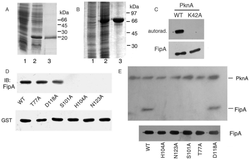
Figure 4. FipA interacts with, and is phosphorylated by PknA in vitro.
A, B. Coomassie blue-stained gels of uninduced (1), or induced (2) cells of E. coli expressing FipA (A), GST-PknA13-273 (B), purified FipA (A, lane 3) and GST-PknA13–273 (B, lane 3). C. Purified FipA was phosphorylated by GST-PknA13–273 or its K42A mutant in vitro in the presence of [γ-32P] ATP followed by autoradiography. D. Recombinant S-tagged FipA or its mutants were incubated with GST-PknA13–273 bound to glutathione-Sepharose beads. Proteins bound to the beads were detected by immunoblotting with anti-FipA antibody. The blot was reprobed with anti-GST antibody. E. Purified FipA or its mutants were phosphorylated by GST-PknA13–273 in vitro in the presence of [γ-32P] ATP followed by autoradiography. The first lane represents autophosphorylated GST-PknA13–273. Blots shown are representative of three separate experiments. Lower blots of panels C and E are Coomassie blue-stained gels showing the levels of FipA.

