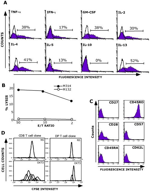
Figure 3. Functional properties of the M314.132 DP T cell clone.
A/ Cytokine production analysis. DP T cell clone was fixed, permeabilized and stained for cytokines following autologous melanoma stimulation. Data are expressed as mean % of intracellular cytokine secreting cells. Open histograms correspond to the analysis of cytokine production by unstimulated M314.132 DP T cell clone (negative control). B/Lysis of the M314 autologous melanoma cell line (closed circles) by M314.132 DP T cell clone. The M132 cell line was used as negative control target (open circles). 51Cr-labeled tumor cells were co-cultured with T cells at various E/T ratios. Chromium release in the supernantants was measured after a 4-h incubation period. C/ Phenotypic characterization of M314 DP T cell clone. D/ Proliferation capacity. CFSE-labeled T cell clones were stimulated with anti-CD3 (OKT3). The CD8 T cell clone used as positive control was obtained by limiting dilution of melanoma specific CD8 T cells. As negative control, T cell clones were maintained in the absence of any stimulation (Not Stimulated: NS).

