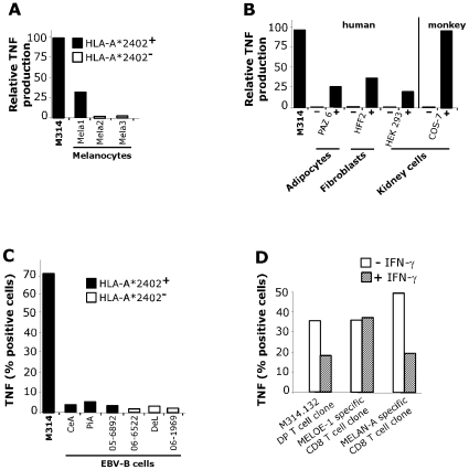
Figure 5. Reactivity of M314.132 DP T cell clone against normal cell lines.
A/ TNF secretion by the M314.132 DP T cell clone in response to melanocytes. 104 DP T cells were added to 3×10⁴ HLA-A*2402 positive or negative melanocytes, and the clone reactivity was assessed by a TNF release assy. B/ TNF secretion by the M314.132 DP T cell clone to HLA-A*2402 transfected (+) or non-transfected (−) normal cells of different origins and/or species. Results are expressed as relative reactivity to the indicated cells in comparison with TNF secretion (100%) induced by M314 autologous melanoma cells. C/ Lack of recognition of HLA-A*2402 EBV-B lymphocytes. DP T cell clone was fixed, permeabilized and stained for cytokines following stimulation with EBV-B cell lines expressing or not HLA-A*2402 molecules. Data are expressed as mean % of intracellular TNF-α secreting cells. D/ TNF response of DP T cell clone toward melanoma cells treated with or without IFN-γ. Melanoma cells were cultured in the presence or absence of 100U/ml rIFN-γ for 15 days. DP T cell clone and two CD8 T cell clones used as controls were fixed, permeabilized and stained for TNF following stimulation with untreated (white bars) or IFN-γ-treated (hatched bars) melanoma cells. Data are expressed as mean % of intracellular TNF-α secreting cells.

