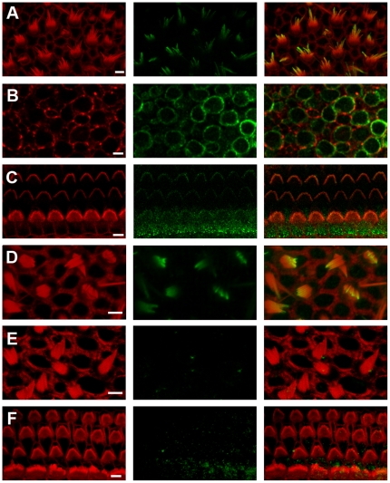
Figure 2. Immunolocalization of HCN subunits in the inner ear.
All panels show confocal images of mouse inner ear epithelia with phalloidin staining shown on the left, HCN1 in the center and the merged image on the right. Phalloidin is shown in red and HCN1 in green. All scale bars indicate 5 µm. (A) Stereociliary bundles of wild-type mouse utricle at P8 stained with an antibody directed against the N-terminus of HCN1. (B) Basolateral hair cell membranes of wild-type mouse utricle stained with the N-terminal HCN1 antibody. (C) Stereociliary bundles of wild-type mouse cochlea harvested from the apex at P8 stained with same N-terminal HCN1 antibody. (D) Confocal image of the stereociliary bundles from a P8 utricle of a HCN1−/− mouse stained the same HCN1 antibody shown in panels A–C. (E) Wild-type utricle focused at the hair bundle level stained with a different antibody that recognizes an epitope in the C-terminus. (F) Wild-type cochlear hair bundles stained with the antibody that recognizes the epitope in the C-terminus.

