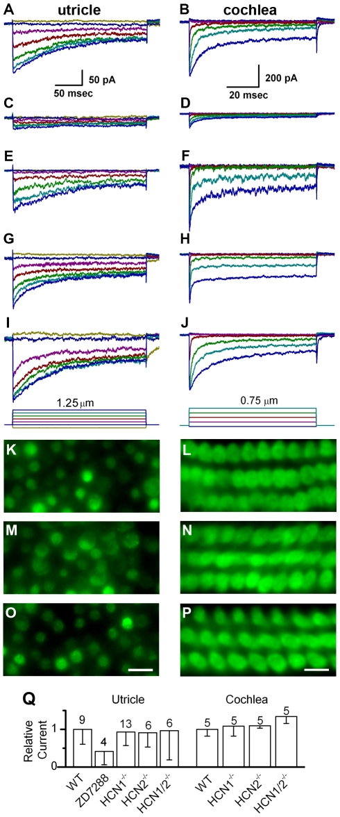
Figure 3. Mechanotransduction in wild-type and HCN-deficient hair cells.
Representative mechanotransduction currents evoked by hair bundle deflections. The left column of data were recorded from mouse utricle type II hair cells at P3 - P6. The data in the right column were recorded from mouse cochlear outer hair cells at P6 - P7. The scale bars at the top apply to all data in the column. The deflection protocol is shown at the bottom of trace panels I and J. Data were recorded under the following conditions: (A & B) wild-type control; (C & D) in the presence of 500 µM ZD7288; (E & F) HCN1−/−; (G & H) HCN2−/−; (I& J) HCN1−/− and HCN2−/−. Panels K-P show fluorescent images of hair cells following application of the transduction channel permeable dye, FM1-43, in utricle (P5 –P7) and organ of Corti (P6 – P9) sensory epithelia under the following conditions: (K & L) HCN1−/−; (M & N) HCN2−/−; (O& P) HCN1−/− and HCN2−/−. Uptake of FM1-43 appeared normal in all tissues examined. The scale bar in panel (O) equals 10 µm and also applies to panels K and M. The scale bar in panel (P) equals 5 µm and also applies to panels L and N. (Q) Summary of transduction currents recorded from vestibular and auditory hair cells. Maximum current amplitudes under each condition were averaged and were normalized relative to wild-type controls. Error bars show standard deviation; the number of samples is indicated above each bar.

