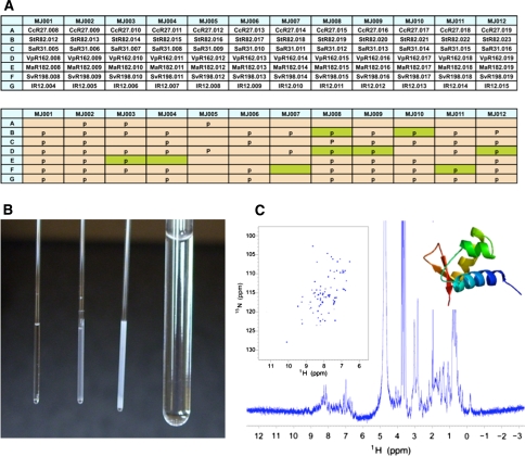
Fig. 6.
a Buffer optimization block of seven NESG protein targets (NESG IDs: CcR27, StR82, SaR31, VpR162, MaR182, SvR196, IR12) in twelve buffers (MJ001 to MJ0012, Supplementary Table S1). Protein signal is detected (pale green) even though the tube shows abundant precipitation indicated by a ‘p’ in the grid. b From the left, 1-mm microtubes showing no or increasing degrees of precipitation of target StR82 in different buffers. Signal is detected in the center tube, but not in the clear (left) or the heavily precipitated (right) microtubes. c The best spectra for pefl from S. typhimurium (NESG target StR82) were recorded at 20°C in 450 mM NaCl at pH 6.5 (buffer MJ008). The insets show the 2D 1H-15N HSQC and the ribbon diagram of the structure solved using optimal conditions (PDB ID: 2JT1)

