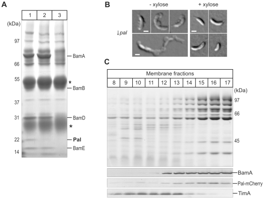
Figure 4. Pal is an essential outer membrane protein, associated with the BAM complex.
(A) BAM complex was immunoprecipitated using BamA antiserum added to outer membrane vesicles that were solubilised with 0.75% (w/v) (Lane 1) and 2.25% (w/v) (Lane 2) dodecyl-maltoside. Immunoprecipitate obtained with preimmune serum was loaded in Lane 3. Asterisks indicate the IgG heavy and light chains, with the migration positions of the molecular markers shown in kDa. (B) Cells with the pal gene under the control of a xylose-inducible promoter (↓ pal) were grown in the presence (right montage) and absence (left montage) of xylose (0.3% [w/v]) for 10 hrs. Outer membrane blebs that form predominantly from the division site or cell poles are evident only in the Pal-depleted cells. Scale bars (white) represent 1 micrometer. (C) Membranes were fractionated on sucrose gradient and analysed by SDS-PAGE. Coomassie Brilliant Blue staining (upper panel) reveals separation of the membrane protein profiles and immunoblotting (lower panel) for the inner membrane protein TimA and the outer membrane protein BamA, and the mCherry epitope to determine the location of Pal.

