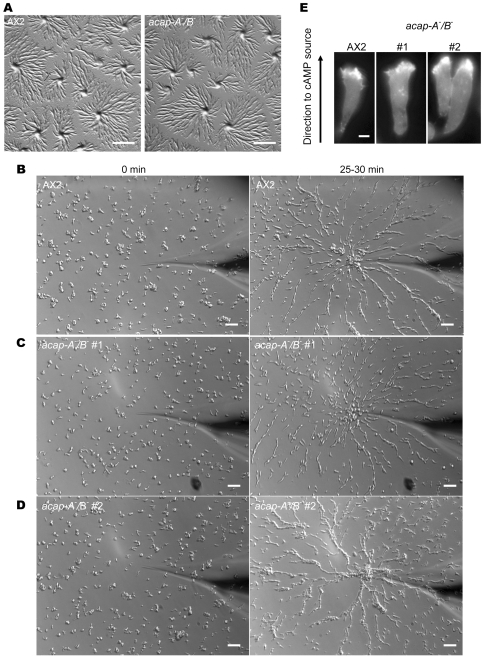
Figure 6. ACAP-A/B are dispensable for chemotaxis and streaming in Dictyostelium.
A, images were taken 6 h after cell were plated on non-nutrient agar as described in Fig. 3. B–D, acap-A−/B − cells migrated towards exogenous cAMP and formed streams. Wild-type (B) or acap-A−/B − cells (C, D) were differentiated with cAMP pulses for 5–6 h and subjected to a point source of 1 µM cAMP. Images were taken every 10 sec by time lapse microscopy as described in “Methods.” Data shown were representative of at least five experiments with two independent clones of acap-A−/B − cells. Also see Videos S1–S4. E, representative images of wild-type AX2 or acap-A−/B − cells expressing mRFPmars-LimEΔcoil during chemotaxis in a gradient of cAMP. Bars, 1 mm (A); 25 µm (B–D); 6 µm (E).

