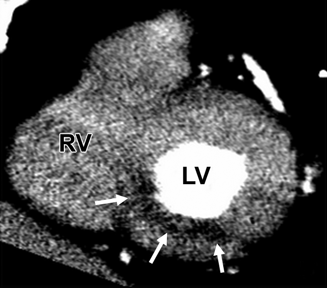
Figure 5b:

Images of 67-year-old man with substernal chest pain and 4-mm ST segment elevation on echocardiogram. (a) Coronary angiogram shows total occlusion of proximal right coronary artery (arrow) that was successfully stented. (b) Multidetector CT study 5 days later reveals area of hypoperfusion (arrows) in inferoseptal and inferior walls, representing MI. Patient also underwent SPECT myocardial perfusion imaging at rest for determination of infarct size on same day of CT. (c) Short-axis and (d) vertical long-axis images did not reveal PD.
