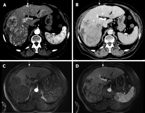
Figure 4.
82-year-old man with biopsy-proven HCC. Detection of an additional tumour nodule by MDCT. The contrast-enhanced arterial phase MDCT demonstrates large tumours in the right liver lobe and one additional hypervascularized nodule in segment 4 (A, arrow) but not in the portal venous phase (B, arrow). Contrast-enhanced MRI depicts the large tumours in the right liver lobe but not in segment 4 (arrows) in early arterial phase (C) and portal venous phase (D).

