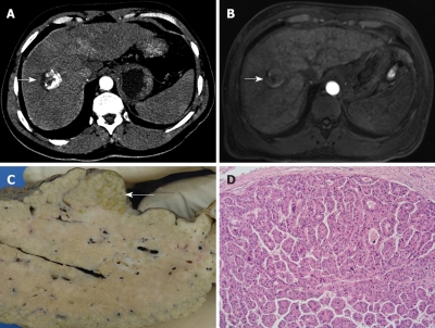
Figure 6.
54-year-old man with biopsy-proven HCC. False-negative finding in the two modalities. Contrast-enhanced early arterial and portal venous phase MDCT (A) and arterial and portal venous phase MRI (B) detected a 3 cm tumour in the right liver lobe (A, B, arrows) but failed to detect another tumour nodule at the posterior surface of the left liver lobe. The explanted liver specimen clearly depicts this additional 2 cm tumour nodule on gross-sectional pathology (C, arrow) and histology (D, 10 × magnification, HE staining).

