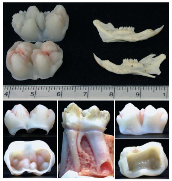
Fig. 1. Tooth size and stage of development.

The developing molars used in our studies are just finishing crown formation, but have not yet started root development. They are about 2 cm in mesialdistal dimension, compared with an adult rat jaw, which is about 3 cm long. On the lower left are two views of the porcine 2nd molar from a 5 to 6 month old pig. Note the open pulp chamber allows easy removal of the pulp at the time of extraction, minimizing contamination by blood components. This contrasts with the erupted 1st molar (center and right). The crown was cut off with a rotary instrument and cleaned. Note the dentin in the 6 month 2nd molar (left) is at an early stage of development and the ceiling of the pulp chamber shows the forms of the cusps, while the 6 month 1st molar has a nearly smooth pulp ceiling. Despite being at an early stage of development, the dentin proteins extracted from the 2nd molars have already experienced extensive proteolysis.
