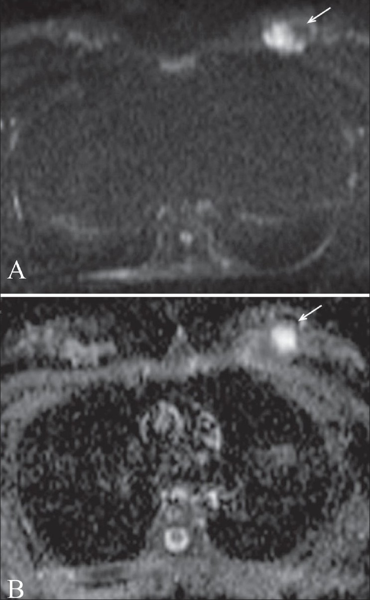
Figure 3.

(A,B): False-negative result in a patient with invasive ductal carcinoma. Diffusion-weighted image (A) at a b value of 1000 shows a left breast mass (arrow) with solid and cystic components. This lesion was proven to be malignant after surgery. Apparent diffusion coefficient (ADC) mapping (B) reveals restricted diffusion (arrow) in the solid component of the mass. The region of interest containing the solid and cystic component reveals an ADC value of 1.5 mm2/s
