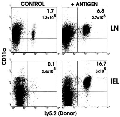
Figure 1.
Appearance of CD8 T cells in the intestinal mucosa after activation of transferred T cells. The adoptive transfer method was modified from Kearney et al. (17). C57BL/6-Ly5.2 OT-I T cells (1 × 10⁷) were injected i.v. into C57BL/6-Ly5.1 mice. Two days later 5 mg of ovalbumin (+ antigen) was administered i.p. Three days later cells were isolated and analyzed for the presence of transferred cells (Ly5.2+) and CD11a expression by fluorescence flow cytometry. Total donor cell numbers were calculated by multiplying the number of Ly5.2+ cells by the total number of LN cells isolated. This experiment has been performed at least 10 times with similar results.

