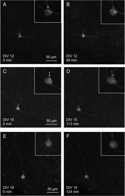
Figure 4.
Multi-day imaging
A neuron transfected with a synaptophysin-CFP plasmid was imaged on 3 different days. The cell was transfected by electroporation on DIV 8. A, B The cell is shown at the beginning (TP 1) and at the end (TP 30) of the first imaging session on DIV 12. The two 3D-images are taken 48 min apart. Inserts show the cell body containing synaptophysin puncta. C, D, E, F The next two imaging days were DIV 15 (D, E) and DIV 19 (G, H). The cell could be identified by its three dendrites. The cell appears healthy, as the synaptophysin puncta are moving about in the cell body and along the dendrites (see movie in supplemental data, Fig. 4s). The inserts show the cell bodies at the respective time points, which show no sign of blebbing.

