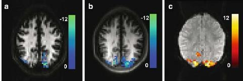
Fig. 1.

Typical EPI (a) and HASTE (b) VASO images with activation overlays (t scores, P < 0.0004). Activation regions detected in the HASTE data are considerably larger, suggesting superior functional sensitivity.As a result, the activation pattern of HASTE more closely resembles that of the BOLD measurements (c). The use of pure spin echoes results in nearly factor 1.7 higher grey matter intensity in the HASTE images as compared to the EPI images
