
Figure 4.
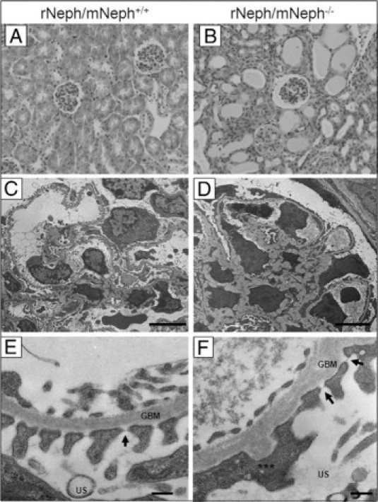
The kidneys from 6-week-old rescued rNeph/mNeph−/− mice showed proteinuria-associated histological abnormalities and a variety of podocyte damage. A and B: H&E staining of paraffin-embedded kidney sections from rNeph/mNeph+/+ and rNeph/mNeph−/− mice (magnification ×200). C–F: Electron microscopy showed a variety of morphological changes such as a foot process effacement, mesangial expansion, and glomerular basement membrane thickening in podocytes of the rescued rNeph/mNeph−/− mice, as compared with rNeph/mNeph+/+ littermate controls. Despite of foot process effacement, slit diaphragm structures were still observed in rNeph/mNeph−/− mice (F). Arrows indicate still existing slit diaphragms (SDs). Asterisks indicate area were partial foot process effacement can be seen. Glomerular basement membrane (GBM), urinary space (US). Scale bars: 5 μm (C and D); 200 nm (E and F).