
Figure 4.
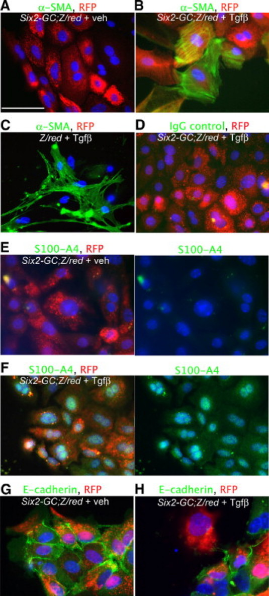
Cultured tubule cells from kidneys of Six2-GC; Z/Red mice, co-express αSMA and S100A4 cultured in the presence of transforming growth factor-β1. A: Day 10 cultured Six2-GC; Z/Red tubule cells fluoresce red from RFP expression, but do not label for αSMA. Scale bar = 50 μm. B: Day 10 cultured Six2-GC; Z/Red tubule cells grown in the presence of TGFβ1 for 4 days fluoresce red with RFP and co-label for αSMA (green). C: Day 10 cultured Z/Red tubule cells grown in the presence of TGFβ1 for 4 days show no evidence of RFP expression but co-label with αSMA (green). D: Day 10 cultured Six2-GC; Z/Red tubule cells grown in the presence of TGF1 for 4 days fluoresce red with RFP show no evidence of non-specific binding with irrelevant IgG control primary antibodies. E: Split panel images (right shows green channel only) Day 10 cultured Six2-GC; Z/Red tubule cells fluoresce red with RFP and do not label for S100A4. F: Split panel images showing Day 10 cultured Six2-GC; Z/Red tubule cells grown in the presence of TGFβ1 for 4 days fluoresce red with RFP and co-label for S100A4 (green) in the cytoplasm and nucleus. G: Day 10 cultured Six2-GC; Z/Red tubule cells fluoresce red with RFP and label for E-cadherin (green). H: Day 10 cultured Six2-GC; Z/Red tubule cells grown in the presence of TGFβ1 for 4 days also fluoresce red with RFP and co-label for E cadherin (green), however some cells lose E-cadherin expression.