
Figure 3.
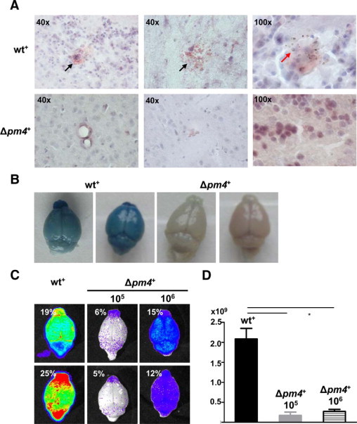
Analysis of cerebral complications in mice infected with wt+ and Δpm4+ parasites. A: Histological analysis of the brains of C57BL/6 mice with asynchronous infections at day 7 after injection of wt+ or Δpm4+ parasites. Longitudinal sections of the brain, stained with H&E, show extensive hemorrhagic areas in wt+-infected animals (black arrows), which are absent in Δpm4+-infected mice. In brains of wt+-infected mice, infected erythrocytes, recognized by pigment granules, were found as infiltrates in brain tissue (red arrow). B: Representative digital images of Evans Blue dye extrusion analysis of brains of wt+ or Δpm4+-infected C57BL/6 mice at day 7 postinfection. The blue staining of the brains shows vascular leakage in wt+-infected animals, which is absent in the brains of Δpm4+-infected mice. C: Bioluminescent images of brain isolated from C57BL/6 mice at day 7 after infection with 105 wt+ or 105 to 106 Δpm4+ parasites showing a significant higher parasite load (see D) in brains of wt+ than in brains of Δpm4+-infected mice as a result of differences in accumulation of infected erythrocytes in brain tissue. Percentages shown represent the parasitemias at the time of collection of the brains. D: Differences in luminescence signal between the brains of mice infected with either wt+ or Δpm4+ parasites (n = 6) showing the differences in parasite load. *P < 0.05.