
Figure 6.
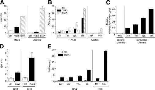
Antigen-specific stimulation of CHS effector T cells from sensitized mice induces their OPN secretion. On day 0, C57BL/6 mice were sensitized on the abdomen with 7% TNCB and challenged with 1% TNCB on both ears on day 5. Twenty-four hours after challenge, lymph node single-cell suspensions were prepared (A–C). Lymph node cells from challenged mice were stimulated with untreated or TNBS-loaded splenic cells or concavalin A (ConA) was added to untreated cells. Proliferation was measured by [3H]thymidine uptake (A), secretion of OPN was detected by ELISA (B), and OPN mRNA expression was analyzed by real-time PCR. Data are given as ratio of OPN mRNA to glyceraldehyde-3-phosphate dehydrogenase housekeeping gene (GAPDH) (C). D and E: CD4+ and CD8+ T cells were isolated by MACS-negative depletion. Cells were cultured with 30-Gy irradiated, TNBS-loaded spleen cells or unloaded spleen cells as controls (Ctrl). Proliferation was measured by [3H]thymidine uptake (D), and secretion of OPN was detected by ELISA (E). The data represent the results of three independent experiments.