
Figure 2.
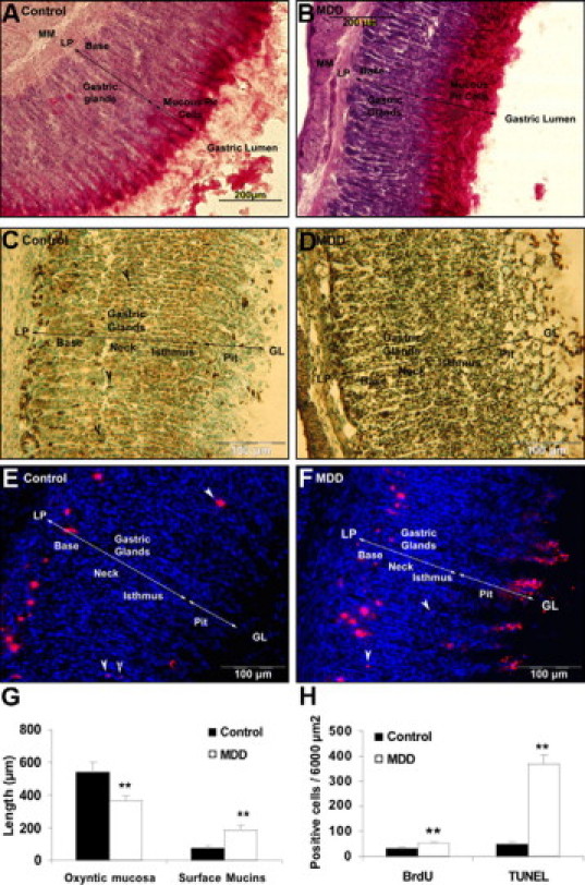
Immunohistological examination of the gastric oxyntic mucosa in control (A, C, E) and MDD rats (B, D, F) at weaning. Mucous cells labeling by PAS staining (A, B). Apoptosis detection by TUNEL assay: in control, pit cells, some neck cells (black arrowheads) and cells from the base region were TUNEL-positive (C), whereas in MDD apoptotic cells were found throughout the mucosa (D). BrdU labeling of proliferative cells: In control rats, newly proliferating cells were located in the isthmus region (E, white arrowheads) and migrated to the base, whereas in MDD pups, BrdU-positive cells were mainly located from the neck to the pit region (F, white arrows); thickness of the total gastric oxyntic mucosa and of the surface mucin staining (G); quantification of proliferative (BrdU positive) and apoptotic (TUNEL) cells in the gastric oxyntic mucosa (H). MM, muscularis mucosa; LP, lamina propria; GL, gastric lumen. **P < 0.01.