
Figure 3.
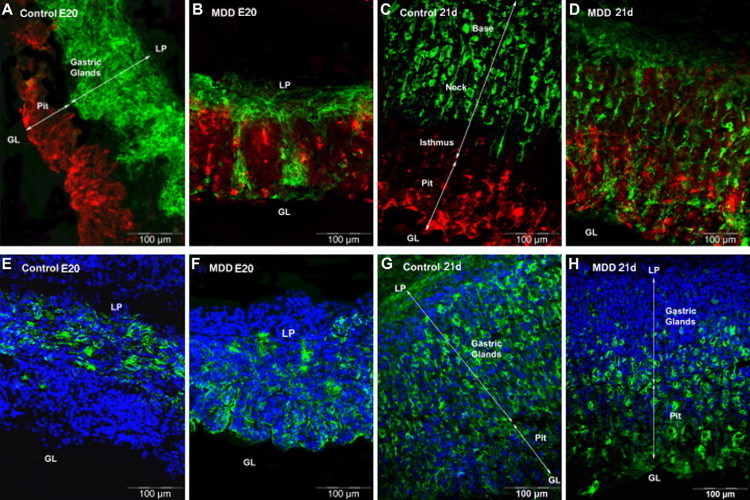
Immunohistological localization of specific cell type among the gastric glands in control and MDD E20 fetuses (four left panels) and rats at weaning (four right panels). A–D: Mucus-secreting pit cells labeled with UEA1 lectin are colored in red, whereas mucus-secreting neck cells labeled with GSII lectin are colored in green. E–H: Parietal cells are colored in green. LP, lamina propria; GL, gastric lumen.