
Figure 2.
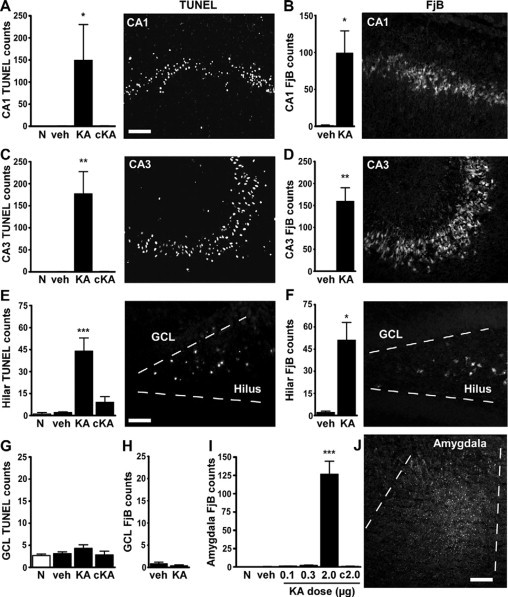
Permanent hippocampal damage 72 hours after neonatal status epilepticus in P10 rats. A–H: Quantification of ipsilateral TUNEL and FjB counts 72 hours after injection of vehicle (veh) or 2 μg of KA compared with age-matched naive (N) pups for CA1 (A and B), CA3 (C and D), hilus (E and F), and dentate granule cell layer (GCL) (G and H). Data from contralateral (c) fields are shown for 2-μg injected pups. Panels to the right of graphs are representative field views of TUNEL and FjB staining from 2-μg KA-injected pups at 72 hours. I: Quantification of FjB counts within the amygdala at the injection site for each dose tested. J: Representative photomicrograph of FjB staining within the ipsilateral amygdala at P13 in a 2-μg injected pup. Data are from n = 3–4 rats per group. ∗P < 0.05, ∗∗P < 0.01, and ∗∗∗P < 0.001 compared with vehicle and naive pups. Scale bar in A–D, 100 μm; E and F, 50 μm; and J, 150 μm.