
Figure 6.
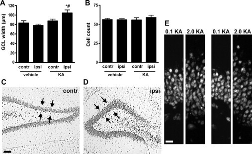
Granule cell layer widening at P55/65 following neonatal status epilepticus. A: Graph showing granule cell layer widths. Significant widening was detected in ipsilateral (ipsi) hippocampus of 2-μg KA-injected pups by adulthood (n = 4–6 per group). ∗P < 0.05 versus ipsilateral side of vehicle control; #P < 0.05 versus contralateral (contr) side of KA injected. B: Graph of granule cell layer counts from the same area show no differences between groups. C and D: Representative photomicrographs of the contralateral and ipsilateral dentate gyrus of a status epilepticus rat at P55 with widening of layer on ipsilateral side. Arrows highlight area of comparison. E: Representative photomicrographs of the upper blade of the ipsilateral dentate granule cell layer from 0.1- and 2-μg KA-injected rats at P55 at two different positions. Note difference in layer width despite numbers of cells being comparable. Scale bars in C, 100 μm; in E, 15 μm.