
Figure 2.
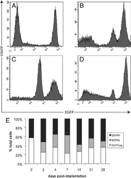
Myeloid cell recruitment to the peritoneal cavity after foreign object implantation. Representative FACS profiles showing EGFP expression by peritoneal exudate cells before (A) and at days 2 (B), 7 (C), or 28 (D) after implantation of cubes of boiled egg white in the peritoneal cavity. E: Histogram shows the proportion of EGFP−, EGFPlo, and EGFPhi cells in the peritoneal cavity before and after implantation. The EGFPlo subset was observed from day 2 onward. Results are expressed as the mean % of total cells (n = 9 mice for each time point from three independent experiments).