
Figure 4.
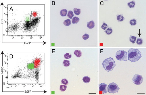
Morphological analysis of peritoneal lavage cells. Cells from the peritoneal cavity at day 2 after implantation stained for Gr1 (A) or F4/80 (D). Morphological analysis (Diff-Quik) of FACS-sorted EGFPloGr1lo (green) and EGFPhiGr1hi (red) subsets shows neutrophils (B) and polymorphonuclear leukocytes and mononuclear cells with ring-shaped nuclei (arrow) (C), respectively. FACS-sorted EGFPloF4/80lo cells were monocytes and neutrophils (E), whereas the EGFPhiF4/80hi subset comprised macrophages (F). Scale bars = 10 μm.