
Figure 2.
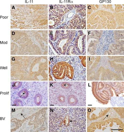
Localization of the site of expression of IL-11, IL-11Rα, and GP130 in endometrial adenocarcinomas and normal proliferative endometrium. The site of expression of IL-11 (A, D, G, J, and M), IL-11Rα (B, E, H, K, and N), and GP130 (C, F, I, L, and O) in samples of poorly (poor, A–C, representative sample 2; Table 1), moderately (mod, D–F; representative sample 15; Table 1), and well-differentiated (G–I, representative sample 26; Table 1) endometrial adenocarcinomas and proliferative-phase endometrium (J–L, representative sample 2; Table 2) by serial section. Intense immunoreactivity as indicated by the brown 3,3′-diaminobenzidine staining was observed to be localized to the glandular epithelial and vascular compartment (BV, M–O as indicated by the arrow, representative sample 29; Table 1) with diffuse stromal immunoreactivity in all tissue sections with no discernable difference in staining pattern observed within each grade or stage of adenocarcinoma. IL-11 and IL-11Rα displayed distinct immunoreactivity on the apical surface in proliferative-phase endometrium (black arrow for IL-11 and IL-11Rα, J and K, respectively) with lesser punctuate staining on the basal (white arrow, for IL-11, J) and lateral surfaces. Control sections were negative for immunoreactivity (Inset, representative serial section of L, representative sample 2; Table 2). Scale bar = 50 μmol/L.