
Figure 3.
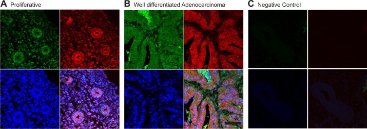
Colocalization of expression of FP receptor, IL-11, and IL-11Rα in well-differentiated endometrial adenocarcinoma and proliferative-phase endometrium. Colocalization of the expression of FP receptor (A and B, lower left panel, blue channel), IL-11Rα (A and B, upper right panel, red channel), and IL-11 protein (A and B, upper left panel, green channel) colocalized (A and B, lower right panel, merged purple channel) in the glandular epithelium and vascular endothelium in proliferative-phase endometrium (A, representative sample 9; Table 2) and well-differentiated endometrial adenocarcinoma (B, representative sample 27; Table 1) by immunofluorescence. The control section C (representative of serial section of representative sample 9; Table 2) was negative for immunoreactivity for all three markers. Scale bar = 50 μmol/L.