
Figure 6.
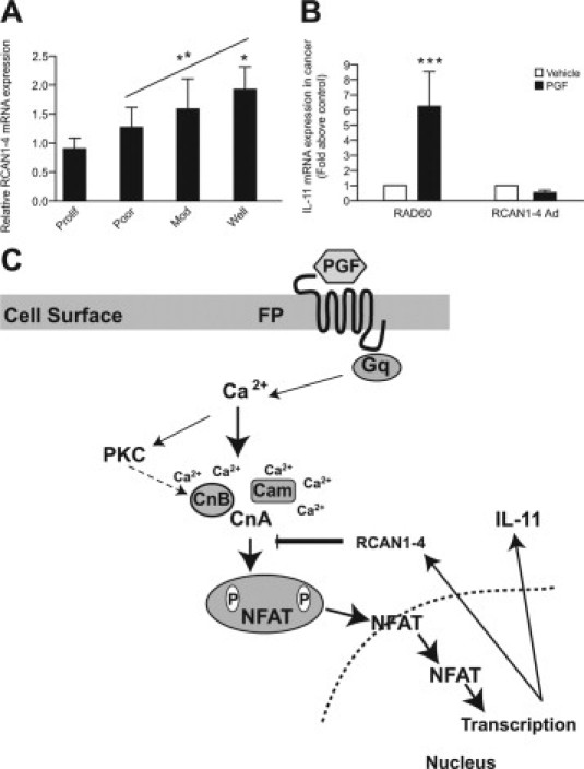
RCAN1-4 expression in endometrial tissues. A: Relative mRNA expression of RCAN1-4 in normal proliferative-phase endometrium (n = 10) and poorly (n = 10), moderately (n = 10), and well-differentiated (n = 10) endometrial adenocarcinoma as determined by real-time quantitative RT-PCR analysis. Data are represented as mean ± SEM *P < 0.03 for well-differentiated adenocarcinoma compared with proliferative-phase endometrium ANOVA posttest for linear trend between poor, moderate, and well-differentiated adenocarcinomas; **P = 0.002. B: Poorly differentiated endometrial adenocarcinomas explants (samples 1 to 4; Table 1) were infected with either RAD60 control adenovirus or RCAN1-4 adenovirus for 24 hours and then stimulated with vehicle or 100 nmol/L PGF2α for 24 hours and subjected to quantitative RT-PCR analysis for IL-11 mRNA expression. Data are represented as mean ± SEM ***P < 0.003 for PGF treatment compared with vehicle control treatment. C: A schematic summary. PGF2α-FP receptor activation in endometrial adenocarcinoma cells promotes the induction of RCAN1-4 and IL-11 via the Gq-phospholipase C-PKC-calcium (Ca2+)-calcineurin-NFAT cascade. RCAN1-4 is temporally activated in a reciprocal manner to IL-11 by PGF-FP receptor signaling and acts as a negative regulator of the calcineurin pathway to regulate IL-11 expression.