
Figure 5.
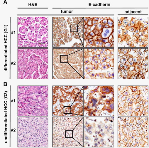
Poorly differentiated human HCC show cytoplasmic dislocation or loss of E-cadherin expression at plasma membrane, suggesting EMT. Human HCC samples were stained with H&E or anti-E-cadherin (E-cadherin) antibody. Two representative examples from both well-differentiated HCC (histological grading G1) (A) and poorly differentiated HCC tissues (histological grading G3) (B) are shown. The left panels show H&E staining. Black boxes in middle panels are regions depicted at fourfold magnification in right panels. E-cadherin is expressed at cell borders in HCC with G1 grade, whereas poorly differentiated HCC with G3 grade show either cytoplasmic delocalization or loss of E-cadherin. Hepatocytes of peritumoral tissues (adjacent) show plasma membrane-localized E-cadherin (right panels). Scale bar = 50 μm.