
Figure 8.
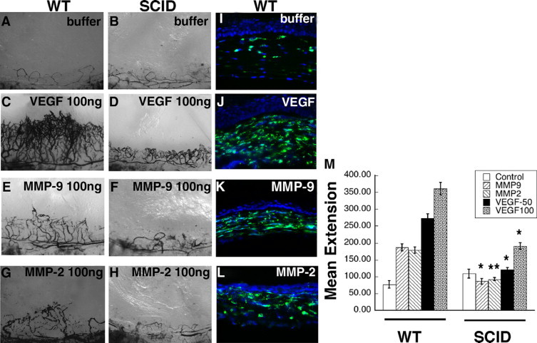
VEGF- and active MMP-induced corneal neovascularization is mediated by inflammatory cells. Representative photographs of mouse corneas (n = 4 for each pellet) at 7 days following implantation of pellets in wild-type (WT) mice containing buffer (A), VEGF (100 ng, C), active MMP-9 (100 ng, E), active MMP-2 (100 ng, G), and in SCID mice containing buffer (B), VEGF (100 ng, D), active MMP-9 (100 ng, F), and active MMP-2 (100 ng, H). Immunohistochemical staining of CD11b in corneas of wild-type mice 24 hours following implantation with buffer (I), VEGF (50 ng, J), active MMP-9 (100 ng, K), and active MMP-2 (100 ng, L). M: Mean vessel extension was calculated as described in Materials and Methods. **P = 0.06, *P < 0.005.