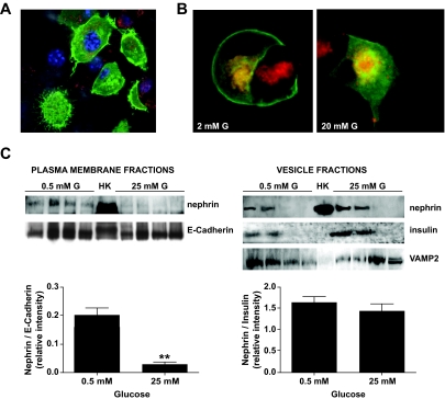
FIG. 6.
Nephrin endocytosis occurs upon glucose stimulation. A: GFP-nephrin–transfected into MIN6 cells localizes both at the plasma membrane and in the cytoplasm. B: MIN6 cells starved in 2 mmol/l glucose revealed that nephrin is predominantly localized to the plasma membrane and only partially to FM6-64–stained vesicles (red). Upon stimulation with 20 mmol/l glucose, nephrin disappears from the plasma membrane and localizes solely to the endocytosed vesicles compartment. C: Plasma membrane and vesicle fractions were collected from MIN6 cells cultured in either 0.5 or 25 mmol/l glucose for 30 min. Human kidney (HK) was used as positive control for nephrin staining. While in 0.5 mmol/l glucose, nephrin was present in both plasma membrane (E-Cadherin positive) fractions and insulin vesicle fractions (insulin positive and VAMP2 positive); in 25 mmol/l glucose, nephrin almost disappeared from the plasma membrane. Shown are a representative blot analysis and the bar graph representation of the nephrin–to–E-cadherin ratios as well as the nephrin-to-insulin ratios (mean and SD of three independent experiments). **P < 0.01. (A high-quality color digital representation of this figure is available in the online issue.)

