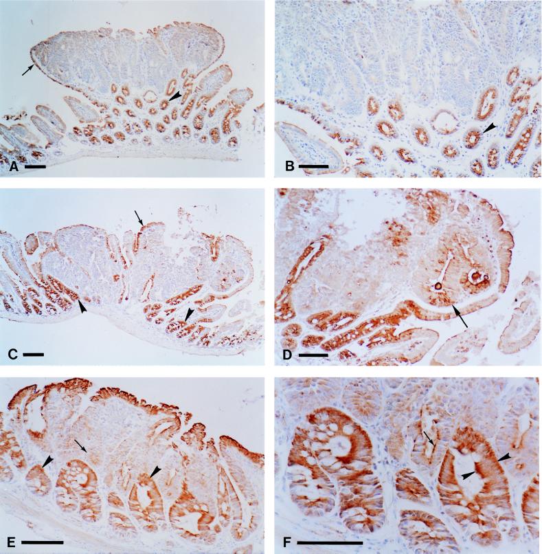
Figure 2.
Analysis of Apc expression in tumors from the small intestine of Min/+ mice. Periodate-lysine-paraformaldehyde-fixed tissues were stained with anti-Apc antibodies as described in Materials and Methods. The tumors in A, B, E, and F were classified by quantitative PCR as showing LOH, whereas the tumor in C and D maintained heterozygosity at Apc. A tumor from an untreated B6 Min/+ mouse stained with the 3122 antibody is shown at low (A) and high (B) magnification. Apc staining is observed in normal crypts (A, arrowhead) and in the normal cells that encapsulate the tumor (A, arrow). Note the strong Apc staining in the apical cytoplasm in the cells from the normal crypts (B, arrowhead). A tumor from an ENU-treated AKR Min/+ mouse stained with 3122 is shown in C and D. Apc staining is seen in normal crypts (C, arrowheads) and in the cells that encapsulate the tumor (C, arrow). A different, serial section of a region of the tumor in C is shown at higher magnification in D. Low levels of staining are observed in some of the tumor cells (D, arrow). A tumor from an untreated AKR Min/+ mouse stained with the APC2 antibody is shown at low (E) and high (F) magnification. Apc staining is much stronger in the cells of the normal crypts (E, arrowheads) relative to the tumor cells (E, arrow). Staining also is seen in the normal cells that encapsulate the tumor. Note the apical and strong basal cytoplasmic staining in the normal crypts (F, arrowheads, compare with B). Some residual staining can be seen in the apical cytoplasm of some of the tumor cells (F, arrow). (Bars, 100 μm for A, C, and E; 75 μm for B, D, and F.)

