
Figure 1. P7 Dizocilpine Increases Caspase-3 Cleavage.
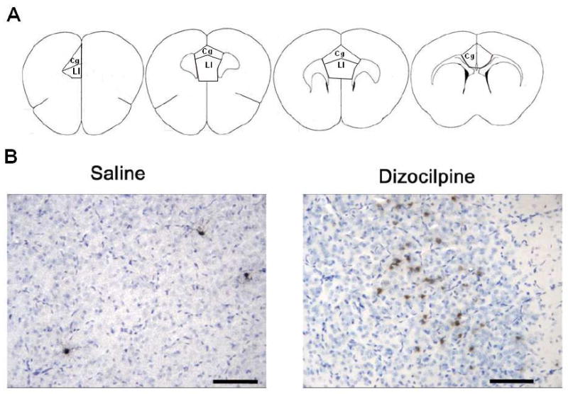
(A.) Diagrams of coronal brain sections between bregma +1.98 to bregma +1.10 that identify the areas of mouse PFC quantified for cleaved caspase-3 immunohistochemistry (Franklin and Paxinos, 2001). Briefly, the dorsal boundary of the anterior cingulate was defined as the diagonal parallel to the dorso-lateral curvature of the fmi (arrows), beginning at the medial peak of the fmi to the medial pial surface. The ventral boundary of the anterior cingulate cortex was defined as a diagonal parallel to the dorsal boundary, beginning one fourth of the total medial length of the fmi, ventral from the peak of the fmi, across to the medial pial surface. (B.) Cleaved Caspase-3+IR. Mice were treated on postnatal day 7 with either saline or dizocilpine (1 mg/kg, 3×24h, i.p.) and assessed for cleaved caspase-3 immunoreactivity (IR) in the prefrontal cortex (PFC) 24 hours after the initial injection. Representative images of cleaved caspase-3 staining with Nissl counterstain (blue) in PFC following either saline or dizocilpine. Note dizocilpine treatment caused an increase in multiple+IR brown cellular profiles indicating cleaved caspase-3+IR. Arrows denote cleaved caspase-3 + IR. Scale bar denotes 50μm.