
Figure 2. Quantification of Cleaved Caspase-3 Immunohistochemistry.
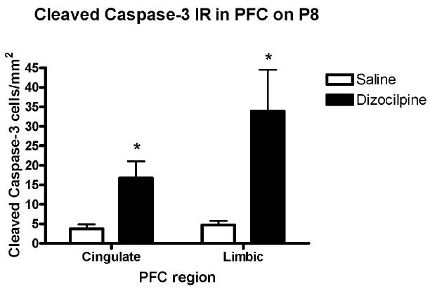
The number of cleaved caspase-3 + immunoreactive (IR) neurons was counted per unit of cortical area as described in the methods. Data shown has been divided into PFC cingulated and PFC limbic as shown in Fig. 1A and described in the methods. Overall PFC cleaved caspase-3+IR cells were increased about 4-7 fold. Within subregions of PFC the anterior cingulate cortex (Cg) increased 4-5 fold and the limbic cortex (LI) increased 6-7 fold (Mean ± standard error, *p<0.03, t-test). Number of mice per treatment group: Saline, n = 5; Dizocilpine, n = 5.