
Figure 1.
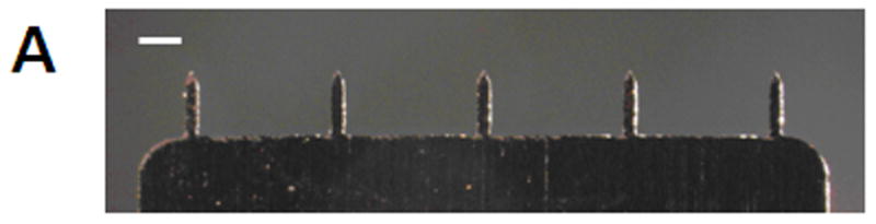
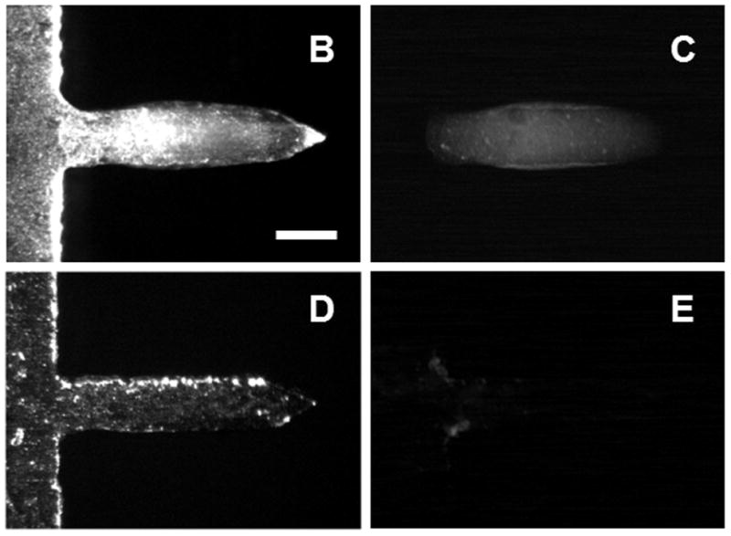
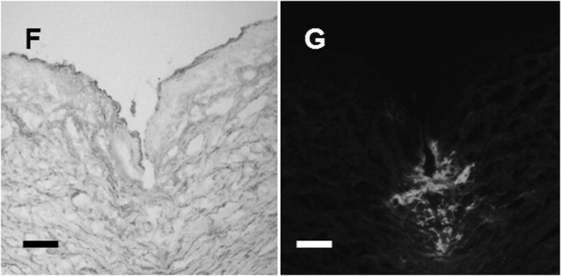
Microneedle coated with influenza vaccine. (A) Image of a 5-microneedle array (scale bar = 500 μm). Bright-field (B, D) and fluorescence (C, E) micrographs of a microneedle coated with red-fluorescent inactivated influenza virus before (B, C) and 10 min after (D, E) insertion into human cadaver skin (scale bar = 200 μm). Histologic section of human cadaver skin fixed after insertion of a vaccine-coated microneedle imaged by (F) bright-field microscopy showing skin deformation and needle track across epidermis and into superficial dermis and (G) fluorescence microscopy showing deposition of red-fluorescent vaccine coating in skin (scale bar = 200 μm).