
Figure 1.
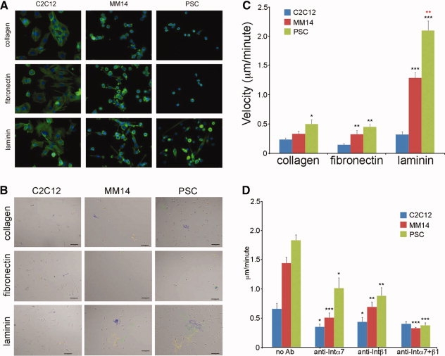
Primary satellite cells and cell lines display differential adhesion and migration on purified extracellular matrix (ECM) substrates; neutralization of α7β1 integrin blocks motility on laminin. (A): Fluorescence micrographs of f-actin (visualized with Alexa 488-phalloidin) and nuclei (visualized with DAPI) of each myogenic cell type plated on different ECM substrates. (B): Final images from representative videos of each cell type on collagen, fibronectin, and laminin; individual cell traces used to calculate average cellular velocity are highlighted in unique colors. Scale bar = 100 μm. Inclusive time on all videos is 14 hours. Movies 1b1, 1b2, and 1b3 correspond to the displayed laminin-adhered cell movies. Number of cells tracked per condition = 12. (C): Primary satellite cells are significantly more motile than cell lines on all substrates; MM14s and primary satellite cells are significantly more motile on laminin than other substrates. Number of cells tracked per condition = 12. (D): Inhibition of integrin α7, integrin β1, or both by neutralizing antibody significantly decreases motility of MM14 and primary satellite cells on laminin. * = p < .05; ** = p < .01; *** = p < .001. Number of cells tracked per condition = 12. Abbreviation: PSC, primary satellite cells.